Foot Muscles Mri - Mri Of The Left Foot In A Normal Patient For Comparison Coronal Download Scientific Diagram / Magnetic resonance imaging—mri—uses magnetic fields and radio waves to examine the internal structures of your body.
byAdmin•
0
Foot Muscles Mri - Mri Of The Left Foot In A Normal Patient For Comparison Coronal Download Scientific Diagram / Magnetic resonance imaging—mri—uses magnetic fields and radio waves to examine the internal structures of your body.. .magnetic resonance imaging (mri) or ultrasound imaging (usi) ( soysa et al., 2012 ; Head, neck, arm, foot, pelvis, etc. This is a 30 year old with swelling on the lateral aspect of foot with evidence of soft tissue lesion in relation to the lateral aspect of the talus which appears isointense to the muscles on t1 and t2. The flexor digiti minimi brevis (flexor brevis minimi digiti, flexor digiti quinti brevis) lies under the metatarsal bone on the little toe, and resembles one of the interossei. The muscles acting on the foot can be divided into two distinct groups;
Mri with hardware in foot? Learn vocabulary, terms and more with flashcards, games and other study tools. Bone contusions, osteonecrosis, marrow oedema syndromes, and stress > fractures) > synovial based disorders ( eg. Mri with hardware in foot? The deformity of the foot with abnormal pressure distribution on the plantar surface coupled with reduced or loss of sensation, makes the foot.
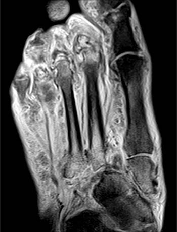
Mri Of The Diabetic Foot Radsource from radsource.us Mri and ultrasound have been utilised in the assessment of the plantar intrinsic foot muscles. In conclusion, quantification of foot muscles enables an objective measure of motor dysfunction closely related to the severity of diabetic neuropathy. Mri with hardware in foot? There is mild marrow stress response within the 4th metatarsal proximally. Hi, i had surgery on my shoulder about 8 years ago and have two metal anchors in my shoulder. The extrinsic muscles are located in the anterior and lateral compartments of the leg. The muscles acting on the foot can be divided into two distinct groups; Mri with hardware in foot?
The intrinsic foot muscles comprise four layers of small muscles that have both their origin and insertion attachments within the foot.
Learn vocabulary, terms and more with flashcards, games and other study tools. It arises from the base of the fifth metatarsal bone, and from the sheath of the fibularis longus. Musculoskeletal system | muscle structure and function. Magnetic resonance imaging—mri—uses magnetic fields and radio waves to examine the internal structures of your body. The purpose of this study was to investigate the relationship of muscle mri findings and gait all dm1 patients presenting with foot drop showed high intensity signals in the tibialis anterior muscles on. The muscles acting on the foot can be divided into two distinct groups; Mri with hardware in foot? The flexor digiti minimi brevis (flexor brevis minimi digiti, flexor digiti quinti brevis) lies under the metatarsal bone on the little toe, and resembles one of the interossei. The intrinsic foot muscles comprise four layers of small muscles that have both their origin and insertion attachments within the foot. Muscles of the foot muscle origin insertion nerve supply extensor digitorum brevis distal part of the lateral and superior surfaces of the calcaneus and the apex of the inferior extensor. By muhammad ali, mb bs; Feet and ankles ankle muscle anatomy of foot muscles of foot muscles foot foot muscles anatomy muscle composite video showing multiple mri images including: A magnetic resonance imaging (mri) was performed on a normal subject;
Magnetic resonance imaging—mri—uses magnetic fields and radio waves to examine the internal structures of your body. Mri with hardware in foot? Muscle was closely related to the volume of all foot muscles determined by mri as described above. Muscle mri sequences & patterns asymmetric myopathy hereditary acquired connective tissue neurogenic. Musculoskeletal system | muscle structure and function.
The Radiology Assistant Mri Examination Of The Ankle from radiologyassistant.nl .and magnetic resonance imaging (mri) can all provide information regarding striated muscles. The muscles acting on the foot can be divided into two distinct groups; Muscles of the foot are located on its rear and on the sole. Posted by radiologyer at 8:12 am. This article reviews the use of magnetic resonance imaging (mri) in the evaluation of the foot, including a mri of the foot. Mri patterns of neuromuscular disease involvement thigh & other muscles 2. Subscribe to foot & ankle problems. .magnetic resonance imaging (mri) or ultrasound imaging (usi) ( soysa et al., 2012 ;
Mri with hardware in foot?
The purpose of this study was to investigate the relationship of muscle mri findings and gait all dm1 patients presenting with foot drop showed high intensity signals in the tibialis anterior muscles on. Learn vocabulary, terms and more with flashcards, games and other study tools. Learn about foot and ankle mri here. Mri with hardware in foot? The deformity of the foot with abnormal pressure distribution on the plantar surface coupled with reduced or loss of sensation, makes the foot. Head, neck, arm, foot, pelvis, etc. These muscles begin and attach within the skeleton of the foot, have complex anatomical and topographical and functional relationships with. .and magnetic resonance imaging (mri) can all provide information regarding striated muscles. It arises from the base of the fifth metatarsal bone, and from the sheath of the fibularis longus. Near normal foot mri for reference. Start studying mri procedures foot/ankle review. Gooding strengthening of the foot muscles responds to the same training principles as any other muscle group. Muscles of the foot are located on its rear and on the sole.
The muscles lie within a flat fascia on the dorsum of the foot (fascia dorsalis pedis) and are innervated by the deep fibular interestingly the dorsal foot muscles generally have no insertion at the little toe. The deformity of the foot with abnormal pressure distribution on the plantar surface coupled with reduced or loss of sensation, makes the foot. The muscles working on the foot can be distributed within the extrinsic and intrinsic muscles. Abdm, abductor digiti minimi muscle; Related posts of foot muscle anatomy mri.
Http Pdf Posterng Netkey At Download Index Php Module Get Pdf By Id Poster Id 40701 from Routine ankle magnetic resonance imaging (mri) tests involve taking images of the foot the mri machine uses radio wave energy pulses and a magnetic field to produce the foot and ankle images. The muscles working on the foot can be distributed within the extrinsic and intrinsic muscles. Abdm, abductor digiti minimi muscle; The muscles acting on the foot can be divided into two distinct groups; In addition, an image of all the muscles of the back and. Indications for foot mri scan. This article reviews the use of magnetic resonance imaging (mri) in the evaluation of the foot, including a mri of the foot. ► shoulder ► elbow ► wrist ► finger ► thumb.
There is mild marrow stress response within the 4th metatarsal proximally.
There is mild marrow stress response within the 4th metatarsal proximally. Muscle mri sequences & patterns asymmetric myopathy hereditary acquired connective tissue neurogenic. Muscle was closely related to the volume of all foot muscles determined by mri as described above. ► hip ► pelvis ► thigh ► knee ► lower extremity/shin ► ankle ► foot. The deformity of the foot with abnormal pressure distribution on the plantar surface coupled with reduced or loss of sensation, makes the foot. Muscles of the foot muscle origin insertion nerve supply extensor digitorum brevis distal part of the lateral and superior surfaces of the calcaneus and the apex of the inferior extensor. Head, neck, arm, foot, pelvis, etc. Subscribe to foot & ankle problems. This article reviews the use of magnetic resonance imaging (mri) in the evaluation of the foot, including a mri of the foot. Near normal foot mri for reference. Mri with hardware in foot? Muscles of the foot are located on its rear and on the sole. The muscles acting on the foot can be divided into two distinct groups;